Body of work
Body of work McGill University
User Tools (skip):
Body of work
The Molson Informatics Project creates art that gets into your mind — and heart, liver and lungs
If you've sat through a first- or second-year medical school lecture, chances are good you've seen the artwork of Joanne Hui, Shie Kasai, Frédéric Léonard, Justin Stephens or Sherwin Tjia without even knowing it. They're the medical illustrators of the Molson Informatics Project, part of the McGill Faculty of Medicine's efforts to bring top-notch visual tools to the classroom.
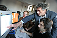
Left to right: Justin Stephens, Dr. David Fleiszer, Frédéric Léonard
Owen Egan
So much medical know-how is easier to comprehend through demonstration than through text, like a Parkinsonian gait, or an irregular heartbeat. When professors supplement words with images and animated sequences during lectures, students will be better able to grasp the medical functioning of the body, whether they learn best spatially or linearly.
Project administrator Nancy Posel says the multimedia learning objects are particularly useful for those conceptual leaps that are difficult to communicate. "There's that moment when you look at the class, and the class looks back at you, and you realize you are not connecting with them completely," Posel laughs.
"The focus of the project is to help enhance understanding."
Joanne Hui
David Fleiszer, a breast and trauma surgeon, is the project's director. He remembers his own student-era problem in understanding the axon's function in nerve disease, and getting it wrong on an exam. The different ways that neurons fire to muscle fibres during disease is complicated to describe. But with a simple animated cartoon, it becomes clear even to a non-scientist.
Back in the eighties, he and his colleagues would pass slides back and forth amongst themselves to accompany their lectures, but the logistics of sharing a single image among busy professionals were time-consuming and unwieldy. At one point, Fleiszer estimated that ten teacher-surgeons had 15,000 slides between them. He organized a single library they could all draw on, but even back then he dreamed of having the technology to digitize all the images for easy access.
Fast forward to the late nineties. With support from the Molson Foundation, he set up a group that doctors could approach to create just the visual tools they need for their lectures.
Shie Kasai
The Molson Project team agreed from the outset that drawings have to be clear, easy to look at and each component must be distinguishable at a glance. It's an improvement over the black marker scribbles that doctors would make on overhead projections in an attempt to show the ebbs and flows of the body's functions. Doctors aren't artists. Leave the drawing of organs and synapses to trained professionals.
The Molson Project's five medical illustrators are Concordia University Fine Arts graduates, chosen by Posel for their artistic skills and fearlessness in the face of science.
They meet with professors (both clinicians and researchers) who go over the curriculum for a particular class and provide them with drawings, textbooks, photos or whatever they need to produce a lecture-ready image or animation. Producing an image can take a few days or many weeks, depending on its complexity and whether it is animated or not. The illustrators often have four or five projects on the go, working with multiple computer screens and drawing tablets.
Fleiszer drops in thrice-weekly to help with any questions they have and give advice. Shie Kasai consults with the doctor on an animation she is working on showing the growth of breast cancer and the surgery required to remove it. This will be used by Fleiszer to demonstrate to cancer patients the procedure they'll undergo, from excising the lymph nodes to where the post-op scars will be. He recommends to Kasai that the lines leading to the labels be altered so they won't be confused with the lymph channels. "Remember, I'll be sitting with a patient who's terrified," he says. Later on, the illustration can be made just a bit more sophisticated so it can be used in a classroom setting.
Justin Stephens
Meanwhile, Joanne Hui is working on a sequence showing the development of a woman's breast from pre-puberty to adulthood and the process of lactation. "It's slow," she says. " You have to think about how you're doing it; if there are errors, if you have to revise, add text, ducts...."
Hui says that the doctors are open-minded and like the change from working with medical students to collaborating with artists and thinking visually. Frédéric Léonard adds, pausing from drawing chromosomal chemical processes, "Doctors tell me it forces them to clarify their ideas."
Each illustrator actively pursues multidisciplinary artistic careers as well. Sherwin Tjia recently took vacation time so he could go on a book tour, reading from his latest work of 1,500 pseudo-haikus to audiences in seven cities over eight days. (McGill folk can see his comic strip, "Pedigree Girls," in The McGill Daily.)
Hui, a painter, and Kasai, a sculptor, were invited to do an installation for Asian Heritage Month in a Mile End library. "It's like having two full-time jobs," Kasai says. She puts in hours at her studio after her workday at McGill. "It takes a lot of time and discipline to keep your art going."
Frédéric Léonard
Léonard paints, as well as being a composer. "He's our sound guy," Hui says of her co-worker, who provided a soundscape for one of her art installations. The illustrators collaborate together in their art, as they do in their office.
The illustrators are exposed to experiences outside the usual realm of the art world, from attending surgery ("It was choreographed like a dance," Hui says) to taking photos of cancer patients who are dying. Some are inspired by the patterns and systems within systems they observe. But not Justin Stephens, who's done a lot of cellular drawings for McGill's histologists. "I try not to let it affect my painting. If anything, my painting influences my drawing here. My girlfriend will come in here to see my work and say, 'That's so you,'" he laughs.
Stephens is currently creating the visuals for case studies that will go on the web. Soon students will be able to visit a virtual hospital, enter a clinic and meet and treat a computer-generated patient by reading files containing the patient's history and chief complaint. The waiting room is modelled after one in the Royal Victoria Hospital, and the virtual patients sitting in the chairs are digitized images of people from the Molson Project's office.
Fleiszer and Posel have big plans for the group's work, such as creating a bank that the 16 Canadian medical schools can draw on. Teachers from across the country can put their best images online. "Instead of 16 mediocre animations of a muscle contraction, there can be one relatively expensive, really well done one," Fleiszer explains.
The online 'waiting room' will be widely accessible to doctors who can input real cases and students who can test their medical acumen against the pros. And if there's dissent over the diagnoses? Fleiszer smiles. "All the better." What matters is that the profession benefits from technology's latest tools and the talents of artists.
For examples of the Molson Informatics Project artists' work, go to a sampler at http://curriculum.mmi.mcgill.ca.